Acute Pain related to Myocardial Infarction

Nursing Care Plan for Acute Pain Myocardial Infarction
Nursing Diagnosis : Acute Pain related to Myocardial Infarction 

Goals and Expected outcomes :
  • Expected loss or uncontrolled chest pain

The expected outcomes:
  • Patients are able to demonstrate the use of relaxation techniques.
  • Patients showed reduced stress, relaxed and easy to move.

Nursing Interventions - Acute Pain related to Myocardial Infarction

Independent
1. Monitor or record the characteristics of the pain, noted the report verbal, nonverbal cues, and the haemodynamic response (grimacing, crying, anxiety, sweating, clutching his chest, rapid breathing, blood pressure / heart frequency change).

Rational: Variations in the appearance and behavior of the patient as pain occurs as the assessment findings. Most patients with Acute Myocardial Infarction looks sick, distraction, and focus on the pain. History of verbal and deeper investigation of the precipitating factors should be delayed until the pain is gone. Breathing may be increased as a result of pain and is associated with anxiety, stress cause temporary loss of catecholamines would increase heart rate and blood pressure.

2. Take a complete picture of the pain of the patient, including the location, intensity (0-10), duration, quality (shallow / spread), and distribution.

Rational:
Pain is a subjective experience and should be described by the patient. Help patients to assess pain by comparing the experience of others.

3. Observations over the previous history of angina, pain resembling angina, or pain in Myocardial Infarction. Discuss family history.

Rational:
Can compare the pain is there from the previous pattern, according to the identification of complications such as widespread infarction, pulmonary embolism, or pericarditis.

4. Instruct patient to report pain immediately.

Rational:
Delays in the reporting of pain, inhibit pain relief and require increased doses of the drug. In addition, severe pain can cause shock by stimulating the sympathetic nervous system, resulting in further damage and interfere with diagnostic and pain relief.

5. Provide a quiet, slow activity, and comfortable action (eg, bed linen dry / not crossed, rubbing his back). Patient approach calmly and with confidence.

Rational:
Lowering external stimuli in which anxiety and heart strain and limited coping abilities and judgment of the current situation.

6. Assist patients in relaxation techniques, eg, deep breathing / slow, behavioral distraction, visualization, imagination guidance.

Rational:
Assist in the reduction in the perception / response to pain. Giving control of the situation, increase positive behavior.

7. Check vital signs before and after drug pemnerian.

Rational:
Hypotension / respiratory depression can occur as a result of the provision. This problem can increase the myocardial damage in the presence of ventricular failure.


Collaboration:

8. Give supplemental oxygen by nasal cannula or mask as indicated.

rational:
Increasing the amount of oxygen available for myocardial usage and also reduce discomfort in relation to tissue ischemia.
12 Nursing Diagnosis for Myocardial Infarction

Myocardial Infarction Nursing Care Plan
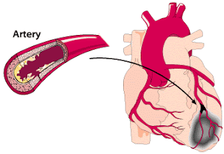
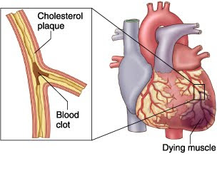
Myocardial infarction (MI) or acute myocardial infarction (AMI) is the death of heart muscle from the sudden blockage of a coronary artery by a blood clot. Myocardial infarction most commonly due to occlusion (blockage) of a coronary artery following the rupture of a vulnerable atherosclerotic plaque, which is an unstable collection of lipids (cholesterol and fatty acids) and white blood cells (especially macrophages) in the wall of an artery. The resulting ischemia (restriction in blood supply) and ensuing oxygen shortage, if left untreated for a sufficient period of time, can cause damage or death (infarction) of heart muscle tissue (myocardium).

Chest pain or pressure is the most common symptom of a heart attack, Myocardial infarction (MI) or heart attack victims may experience a variety of symptoms including:
  • Pain, fullness, and/or squeezing sensation of the chest
  • Jaw pain, toothache, headache
  • Shortness of breath
  • Nausea, vomiting, and/or general epigastric (upper middle abdomen) discomfort
  • Sweating
  • Heartburn and/or indigestion
  • Arm pain (more commonly the left arm, but may be either arm)
  • Upper back pain
  • General malaise (vague feeling of illness)
  • No symptoms (Approximately one quarter of all heart attacks are silent, without chest pain or new symptoms. Silent heart attacks are especially common among patients with diabetes mellitus.)
An MI requires immediate medical attention. Treatment attempts to salvage as much myocardium as possible and to prevent further complications, hence the phrase "time is muscle". Oxygen, aspirin, and nitroglycerin may be administered. Morphine was classically used if nitroglycerin was not effective; however, it may increase mortality in the setting of NSTEMI. A 2009 and 2010 review of high flow oxygen in myocardial infarction found increased mortality and infarct size, calling into question the recommendation about its routine use. Other analgesics such as nitrous oxide are of unknown benefit. Percutaneous coronary intervention (PCI) or fibrinolysis are recommended in those with an STEMI. (wikipedia)


Nursing Care Plan for Myocardial Infarction

12 Nursing Diagnosis for Myocardial Infarction

1. Decreased Cardiac Output
related to:
changes in the frequency of heart rhythm.

2. Impaired Tissue Perfusion
related to:
decrease in cardiac output.

3. Ineffective Airway Clearance
related to:
accumulation of secretions.

4. Ineffective Breathing Pattern
related to:
lung development is not optimal.

5. Impaired Gas Exchange
related to:
pulmonary edema.

6. Acute Pain
relate to:
increase in lactic acid.

7. Fluid Volume Excess
related to:
retention of sodium and water.

8. Imbalanced Nutrition, Less Than Body Requirements
related to:
Inadequate intake.

9. Activity Intolerance
relate to:
imbalance between myocardial oxygen supply and needs.

10. Self-Care Deficit
related to:
physical weakness.

11. Anxiety
related to:
ncaman death.

12. Knowledge Deficit
related to:
lack of information.

Knowledge Deficit Hypertension Nursing Diagnosis Interventions

Nursing Diagnosis and Interventions for Hypertension

Knowledge Deficit related to lack of information about the disease process and self-care.

Purpose:
  • Increased knowledge on the client
Expected outcomes:
  • Clients understand the disease process and treatment.

Nursing Intervention:

1. Assess readiness and barriers to learning, including people nearby.

2. Apply and indicate normal blood pressure limits, explain about hypertension and its effect on the heart, blood vessels, kidneys and brain.

3. Avoid saying normal blood pressure and use the term "well-controlled" when describing the patient's blood pressure patient's blood pressure within normal limits.

Rational:

1. Misconceptions and disprove the diagnosis because of the feeling of well-being has long enjoyed affect the interests of patients and / significant other to study the disease, progression, and prognosis, if the patient does not accept the reality that requires treatment continue, then the behavior changes will not be retained.

2. Provide a basis for understanding the increase in blood pressure and clarify medical terms that are often used, understanding that high blood pressure can occur without symptoms is to allow patients to continue treatment even if you feel healthy.

3. Because treatment for hypertensive patients is through life, then by delivering the idea of ​​"control" will help patients to understand the need for continuing treatment / medication.

10 Nursing Diagnosis for Rheumatic Heart Disease - RHD

Nursing Care Plan for Rheumatic Heart Disease - Nanda Nursing Diagnosis for RHD

Rheumatic heart disease is a condition in which permanent damage to heart valves is caused by rheumatic fever. The heart valve is damaged by a disease process that generally begins with a strep throat caused by bacteria called Streptococcus, and may eventually cause rheumatic fever.

The symptoms of rheumatic fever usually start about one to five weeks after your child has been infected with Streptococcus bacteria. The following are the most common symptoms of rheumatic fever. However, each child may experience symptoms differently.

Symptoms may include:
  • Joint inflammation - including swelling, tenderness, and redness over multiple joints. The joints affected are usually the larger joints in the knees or ankles. The inflammation "moves" from one joint to another over several days.
  • Small nodules or hard, round bumps under the skin.
  • A change in your child's neuromuscular movements (this is usually noted by a change in your child's handwriting and may also include jerky movements).
  • Rash (a pink rash with odd edges that is usually seen on the trunk of the body or arms and legs).
  • Fever.
  • Weight loss.
  • Fatigue.
  • Stomach pains.

The symptoms of rheumatic fever may resemble other conditions or medical problems. Always consult your child's physician for a diagnosis.

Rheumatic fever is uncommon in the US, except in children who have had strep infections that were untreated or inadequately treated. Children ages 5 to 15, particularly if they experience frequent strep throat infections, are most at risk for developing rheumatic fever.

To diagnose this condition, your doctor will ask about recent strep infections, examine your child and use a stethoscope to listen to their heart. In children with rheumatic heart disease, doctors can often hear a heart murmur — the sound of blood moving in the heart in a way that’s not normal.

During the exam, your child’s doctor will look for signs of inflammation in your child’s joints.

The doctor will ask for details about your child’s symptoms, their health history and your family health history. Your doctor may order a throat culture or a blood test to check for strep.

Your child will also need tests that provide information about how their heart looks and works. These may include a chest X-rays or MRI (magnetic resonance imaging) of the heart, echocardiography and electrocardiogram.

Nursing Care Plan for Rheumatic Heart Disease


10 Nursing Diagnosis for Rheumatic Heart Disease - RHD :


1) Decreased Cardiac Output

related to: a disturbance in the closure of the mitral valve (valve stenosis).

2) Ineffective Peripheral Tissue Perfusion

related to: decreased metabolism primarily due to vasoconstriction of peripheral blood vessels.

3) Acute Pain
related to: inflammation of the synovial membrane.

4) Hyperthermia

related to: inflammation of the synovial membrane, and inflammation of the heart valves.

5) Imbalanced Nutrition, Less Than Body Requirements
related to: an increase in stomach acid caused by the sympathetic nervous system compensation.

6) Activity intolerance

related to: muscle weakness, prolonged bed rest or immobilization.

7) Self-Care Deficit

related to: Musculoskeletal Disorders: polyarthritis / arthralgia and therapy bed rest.

8) Impaired Skin Integrity

related to: inflammation of the skin and tissue subcutan.

9) Risk for Impaired Gas Exchange

related to: the accumulation of blood in the lungs due to increased atrial filling.

10) Risk for Injury

related to: involuntary movements, irregular, rapid and muscle weakness / khorea.

Advantages / Benefits of Home Care

Advantages / Benefits of Home Care
Health is the thing that is needed by every human being, it is reflected in the large number of patients who come to health services for treatment and care, they come from many different groups, ranging from high-class economic group, to the down economy.

Home health care is one type of long-term care that can be provided by professionals and non-professionals who have been trained. Home health care, which is one form of health care is a component of a continuous range of health services and comprehensive given to individuals and families in their homes which aims to improve, maintain or restore health and maximizing the level of independence and minimize the consequences of diseases including terminal. Services that meet the needs of individual patients and families, planned, coordinated and provided by the service providers organized to provide home care through staff or contractual arrangements or a combination of both.

Sherwen (1991) defines health care as an integral part of home nursing services performed by nurses to help individuals, families and communities achieve self-reliance in solving health problems that they face. While Stuart (1998) describe the home health care as part of the process of nursing at the hospital, which is a continuation of the repatriation plan (discharge planning), for clients it was time to go home from the hospital. Home care is usually performed by a nurse from the hospital initially, carried out by community nurses where the client is located, or carried out by a special team that handles home care.

Advantages / Benefits of Home Care

The benefits that can be obtained in general from the activities of home care include:
  • Improve promotive, preventive, curative and rehabilitative.
  • Reducing the frequency of hospitalization.
  • Efficiency of time, cost, effort and thought.

The benefits of home care, hospital-based:
  • Direct access to doctors who refer and treat patients and potential patients easier.
  • The carrying capacity of a large organization financially available to help solve temporary cash - flow in the initial phase.
  • Ease / advantage in securing managed care through a process of collaboration and service integration.
  • The need for comprehensive care of patients will be met.
  • Continuity of care and the internal controls of the cost, quality and access to services.
  • Length of Stay will ter-manage more effectively.
  • There are opportunities to increase hospital revenue.

Nursing Care Plan for Atherosclerosis

Nursing Diagnosis for Atherosclerosis
Atherosclerosis (ath-er-o-skler-O-sis) also known as arteriosclerotic vascular disease or ASVD. comes from the Greek words athero - meaning gruel or paste and sclerosis meaning hardness - and is a hardening of the arteries - it is the most common cause of heart disease. Atherosclerosis is a condition in which an artery wall thickens as a result of the accumulation of fatty materials such as cholesterol.

Atherosclerosis usually doesn't cause signs and symptoms until it severely narrows or totally blocks an artery. Many people don't know they have the disease until they have a medical emergency, such as a heart attack or stroke. Some people may have signs and symptoms of the disease. Signs and symptoms will depend on which arteries are affected.

These symptoms take some time to develop, as the disease must progress to the point where an artery is severely narrowed or completely blocked.

Common locations for narrowing and hardening of the arteries to occur include the:
  •     Heart
  •     Brain
  •     Legs, pelvis, or arms
  •     Kidneys.
Symptoms of Atherosclerosis in the Heart
If the arteries that supply the heart with blood (called the coronary arteries) are affected, you may have symptoms that include:
  •     Chest pain or chest discomfort (angina)
  •     Pain in one or both arms, the left shoulder, neck, jaw, or back
  •     Shortness of breath
  •     Dizziness
  •     Faster heartbeats
  •     Nausea (feeling sick to your stomach)
  •     Abnormal heartbeats
  •     Feeling very tired.
In some people, the first symptom is a heart attack. A heart attack occurs when a coronary artery becomes blocked, most commonly by a blood clot.

Causes and Risk Factors of Atherosclerosis

Why does atherosclerosis occur in the coronary arteries of some people but not others? An interplay of many factors including hypertension (high blood pressure), smoking, diabetes, obesity, high cholesterol, family history of heart disease, and a sedentary lifestyle are involved.

Treatment of Atherosclerosis
  • Medication is unsatisfactory for treating atherosclerosis, since the damage has already been done.
  • Anticoagulant drugs have been used to try to minimize secondary clotting and embolus formation.
  • Vasodilator drugs are helpful in providing symptom relief, but are of no curative value.
  • Surgical treatment is available for those unresponsive to medical treatment or in certain high-risk situations.
  • Balloon angioplasty can open up narrowed vessels and promote an improved blood supply.
  • The blood supply to the heart can also be restored by coronary artery bypass surgery.
  • Large atheromatous and calcified arterial obstruction can be removed by endartectomy, and entire segments of diseased peripheral vessels can be replaced by woven plastic tube grafts.

Nursing Care Plan for Atherosclerosis

Physical Examination - Nursing Care Plan for Atherosclerosis

1. Angina Pectoris (chest pain) followed by:
  • The urge to urinate
  • Diaphoresis
  • Nausea
  • Dyspnoea
  • Cold extremities
2. Assess Pain to identify Angina
  • Stable angina is chest pain or discomfort that Usually Occurs with activity or stress. Angina is chest discomfort due to poor blood flow through the blood vessels in the heart.
  • Unstable angina is a condition in roomates your heart does not get enough blood flow and oxygen. It may lead to a heart attack.
  • Nocturnal angina wakes a patient from sleep and may be provoked by vivid dreams. Symptoms are commonest in the early hours of the morning when coronary artery tone is maximal. Often the patient has critical coronary artery disease and hence Usually suffers from exertional angina. Nocturnal angina may be associated with coronary artery spasm - Prinzmetal's angina.
  • Decubitus angina Occurs when the patient lies down. Usually it is a complication of cardiac failure due to the strain on the heart resulting from the Increased intravascular volume. Usually Patients have severe coronary artery disease.
  • Prinzmetal's angina is a form of chest pain, pressure, or tightness (angina) Caused by spasms in the arteries that supply blood to the heart. It is a form of unstable angina, meaning that it Occurs at rest, Often without a predictable pattern. This is in contrast to stable angina, chest pain in roomates Occurs in a predictable pattern during exertion or exercise.

3. Assess the Chest Pain in relation to:
  • Trigger factors in patients, what triggers the onset of pain is to be done before the pain began to occur (eg; smoking, excessive activity, excessive weight diet, emotional stress, sexual activity and drink too cold)
  • Quality pain how (are like a burning sensation, feeling depressed or choke)
  • Location of pain: occurs in substernal or mid anterior chest and around the neck, jaw, or left arm shoulder blades down.
  • Remarkably attacks: mild, moderate or severe.
  • Time; illness duration, frequency.
  • Typical in the attack: a fist over his chest or left arm rub. Pain attacks occur gradually or abruptly for 15 minutes or more.
  • Assess the client's feelings about the conditions and the perceived influence of lifestyle.

Nursing Assessment - Nursing Care Plan for Atherosclerosis

The data should be assessed in patients with atherosclerosis or arteriosclerosis depends on the location affected. When the coronary arteries are exposed to the clinical signs and symptoms according to clinical signs and symptoms of angina pectoris or acute myocardial infarction. When the brain is affected by the clinical signs and symptoms were assessed according to the case of stroke. Angina pectoris, myocardial infarction and stroke will be discussed separately. Nursing assessment will be our focus here is impaired peripheral perfusion of the organs other than those mentioned above.

Subjective data which may occur: sudden pain or felt melancholy, cramps, fatigue or weakness. Pain persists rest, pain, and discomfort, and usually occurs in the distal extremities. Cold feeling or numbness in the extremities due to decreased arterial flow. Assess the level of knowledge of the patient about the treatment of the disease.

Objective data that may be obtained: the affected extremity will look pale when elevated and cyanosis while hanging. The color and temperature of the extremities were recorded. Changes in skin and nails, ulcers, gangrene and muscle atrophy may seem obvious. The nails may thicken and cloudy, shiny skin, atrophy and dry with sparse hair growth. Peripheral pulses can be weakened or lost altogether.

Risk for Impaired Skin Integrity - NCP Guillain-Barre syndrome

Nursing Diagnosis Risk for Impaired Skin Integrity - Nursing Care Plan for Guillain-Barre Syndrome

Guillain-Barre syndrome is a serious disorder that occurs when the body's defense (immune) system mistakenly attacks part of the nervous system. This leads to nerve inflammation that causes muscle weakness and other symptoms.

Symptoms of Guillain-Barre syndrome include:
  •     Numbness or tingling in your hands and feet and sometimes around the mouth and lips.
  •     Muscle weakness in your legs and arms and the sides of your face.
  •     Trouble speaking, chewing, and swallowing.
  •     Not being able to move your eyes.
  •     Back pain.
Symptoms usually start with numbness or tingling in the fingers and toes. Over several days, muscle weakness in the legs and arms develops. After about 4 weeks, most people begin to get better.

You may need to be treated in the hospital for the first few weeks. This is because GBS can be deadly if weakness spreads to muscles that control breathing, heart rate, and blood pressure.

Signs and tests

A history of increasing muscle weakness and paralysis may be a sign of Guillain-Barre syndrome, especially if there was a recent illness.

A medical exam may show muscle weakness and problems with involuntary (autonomic) body functions, such as blood pressure and heart rate. The examination will also show that reflexes, such as the "ankle or knee jerk," are decreased or missing.

There may be signs of decreased breathing caused by paralysis of the breathing muscles.

The following tests may be ordered:
  •     Cerebrospinal fluid sample ("spinal tap")
  •     ECG
  •     Electromyography (EMG) tests the electrical activity in muscles
  •     Nerve conduction velocity test
  •     Pulmonary function tests
Nursing Diagnosis : Risk for Impaired Skin Integrity : dekubitus related to kelemahan otot, paralisis, gangguan sensasi, perubahan nutrisi, inkontinensia.

Expected outcomes:
  • Patients retain the skin remains dry and intact.
  • Maintaining depressed area remains dry and intact, free of pressure sores.
Intervention:

1. Assess motor function and sensation every 4 hours.
R /: muscle paralysis can occur quickly with a pattern that has been rising.

2. Assess the patient's degree of dependence.
R /: To identify patients in need of ADL ability.

3 . Monitor depressed area.
R /: Identifying early signs of pressure sores.

4. Keep the bed, lake remains clean, tight and dry.
R /: Laken, wet, dirty, matted facilitate the occurrence of pressure sores.

5. Monitor intake and output of nutrients.
R /: inadequate nutrition reduce the risk of pressure sores.

6. Perform over the position every 2 hours.
R /: Smooth distressed parts of blood flow.

7. Perform ROM.
R /: Preventing atrophy.

Physical Examination for Meningitis

Physical Examination for Meningitis
Meningitis 

Meningitis is a disease caused by the inflammation of the protective membranes covering the brain and spinal cord known as the meninges. It is most often caused by infection (bacterial, viral, or fungal), but can also be produced by chemical irritation, subarachnoid haemorrhage, cancer and other conditions. The severity of illness and the treatment for meningitis differ depending on the cause. Thus, it is important to know the specific cause of meningitis.

Viral Meningitis
Enteroviruses, the most common cause of viral meningitis, are most often spread from person to person through fecal contamination (which can occur when changing a diaper or using the toilet and not properly washing hands afterwards).

Bacterial Meningitis
Bacterial meningitis is contagious. The bacteria are spread through the exchange of respiratory and throat secretions (i.e., kissing).

The most common symptoms of either form of meningitis include:
  • Fever.
  • Severe and persistent headache.
  • Stiff and painful neck, especially when trying to touch the chin to the chest.
  • Vomiting.
  • Confusion and decreased level of consciousness.
  • Seizures.
Other symptoms of meningitis include:
  • Sluggishness, muscle aches and weakness, and strange feelings (such as tingling) or weakness throughout the body.
  • Eye sensitivity and eye pain from bright lights.
  • Skin rash.
  • Dizzy spells.

Physical Examination for Meningitis

1. Activity / Rest
  • Symptoms: feeling unwell (malaise), limitations posed condition.
  • Signs: Ataxia, problems walking, paralysis, involuntary movement, general weakness, limitations in range of motion.
2. Circulation
  • Symptoms: the history of cardiology, such as endocarditis, some heart diseases Conginetal (brain abscess).
  • Symptoms: increased blood pressure, decreased pulse rate, pulse pressure weight (associated with increased ICP and the influence of the vasomotor center). Tachycardia, distritmia (acute phase) as distrimia sinus (in meningitis)
3. Elimination
  • Signs: The existence of urinary incontinence and retention.
4. Food and Fluids
  • Symptoms: Loss of appetite, difficulty swallowing (acute period)
  • Signs: Anorexia, vomiting, poor skin turgor, dry mucous membranes.
5. Hygiene
  • Signs: Dependence on all the needs of self-care (acute period)
6. Neurosensory
  • Symptoms: headache (may be the first symptom, and usually heavy), paresthesia, feels stiff in all the nerves are affected, loss of sensation (cranial nerve damage). Hyperalgesia / increased sensitivity (minimitis). Seizures arise (minimitis bacteria or brain abscess) disturbances in vision, such as monocular (early phase of multiple infections). Photophobia (on minimtis). Deafness (on minimitis / encephalitis) or maybe hypersensitivity to noise, the hulusinasi smell / touch.
  • Signs:
    • Mental status / level of consciousness; lethargy to severe confusion to coma, delusions and hallucinations / psychosis organic (encephalitis).
    • Memory loss, difficulty in making decisions (can be a symptom of growing hidrosephalus communicant, following bacterial meningitis)
    • Aphasia / difficulty in communicating.
    • Eyes (size / pupil reaction): unisokor or do not respond to light (increased ICP), nystagmus (eyes move continuously).
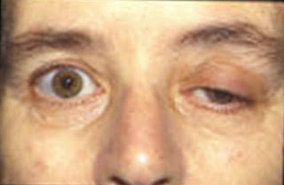
    • Upper eyelid ptosis falling). Characteristic facial (face), changes to the motor and sensory functions (cranial nerves V and VII exposed)
    • Generalized seizures or locally (on the brain abscess). Temporal lobe seizures. Experiencing muscle hypotonia / flaccid paralysis (acute phase of meningitis). Spastic (encephalitis).
    • Hemiparese hemiplegic (meningitis / encephalitis)
    • Brudzinski's sign positive, positive Kernig sign, an indication of meningeal irritation (acute phase)
    • Regiditas face (meningeal irritation)
    • Deep tendon reflexes disturbed, positive Brudzinski
    • Abdominal reflexes decreased.

7. Pain / Leisure
  • Symptoms: headache (throbbing madly, frontal) may be exacerbated by tension neck / back stiffness, pain on ocular movement, throat pain.
  • Signs: Looks kept awake, behavioral distraction / agitated crying / complaining.

8. Breathing
  • Symptoms: A history of sinus or lung infections.
  • Signs: Increased work of breathing (early stage), mental changes (lethargy to coma) and restless.

9. Security
  • Symptoms:
    • A history of upper respiratory tract infection or other infections, including sinus middle ear mastoiditis, dental abscess, abdominal or skin, lumbar function, surgery, fracture of the skull / head injury.
    • Immunizations are just getting under way; exposed to meningitis, exposed to measles, herpes simplex, animal bites, foreign bodies carried away.
    • Impaired vision or hearing
  • Signs:
    • Increased body temperature, diaphoresis, shivering
    • General weakness; flaccid muscle tone or plastic
    • Sensory disturbances.

Ineffective Breathing Pattern - Ineffective Airway Clearance - Impaired Gas Exchange

Nursing Interventions for Guillain-Barre Syndrome

Nursing Care Plan for Guillain-Barre Syndrome

Guillain-Barre syndrome is a severe inflammatory disorder of the peripheral nerves. It is an autoimmune disease, i.e. the immune system that is supposed to attack foreign substances like bacteria; starts attacking cells of own body, in this case the nerves. The immune system produces special molecules, called the antibodies that are mainly responsible for damage to nerve cells in Guillain-Barre Syndrome. A previously healthy person suddenly develops tingling and numbness primarily in the feet which within a couple of weeks spreads through the body to cause loss of muscle control and feeling throughout the body.

Symptoms of Guillain-Barre syndrome :
  • Lack of feeling
  • Weakness or itchiness in arms or legs
  • Possible loss of feeling and movement in the upper body, face, arms and legs.
The symptoms can remain in this phase and can cause little difficulty in walking. However, in some cases the illness can progress resulting in entire paralysis of arms and legs.

Diagnosis

Gullain Barre syndrome is considered to be the most harmful disorder because it attacks the patient suddenly and surprisingly. The patient within weeks reaches the highest level of weakness. In 3rd or 4th weeks of the illness the patients are at their weakest. The recovery period can be varying according to the condition of the patient. It can be few weeks or in some cases a few years.


Nursin g Interventions for Guillain-Barre Syndrome

Ineffective Breathing Pattern, Ineffective Airway Clearance, Impaired Gas Exchange related to respiratory muscle weakness or paralysis, decreased cough reflex, immobilization.

Ineffective Breathing Pattern Definition : The exchange of air inspiration and / or expiration inadequate.

Ineffective Airway Clearance Definition: Inability to clear secretions or obstructions from the respiratory tract to maintain airway patency.

Impaired Gas Exchange Definition : Circumstances where an individual has decreased course of gas (O2 and CO2) that an actual or risk of lung alveoli and the vascular system.

Expected outcomes:
  • Optimal breathing.
  • Normal breath sounds.
  • Patent airway.
  • Blood gas analysis values within normal limits.
Intervention:

1. Monitor the number of respiratory rhythm and depth every 1-4 hours.
R /: Paralysis of breathing can occur 48 hours.

2. Auscultation of breath sounds every every 4 hours.
R /: breath sounds indicate inadequate ventilation.

3. Maintain effective airway, suction and clean the mouth.
R /: a patent airway.

4. Help the patient to cough effectively.
R /: Increase effective airway.

5. Perform chest physiotherapy.
R /: Preventing pneumonia and atelectasis.

6. Collaboration in the provision of oxygenation.
R /: Fulfilling the need of oxygen.

7. Monitor blood gas analysis.
R /: Knowing the changes in oxygen in the blood.

8. Assess the level of consciousness and skin tone.
R /: Changes in blood gas analysis will affect the level of consciousness and skin tone.

Priority Nursing Diagnosis for Hypertension

Priority Nursing Diagnosis for Hypertension


Hypertension is among the world's famous disease. Hypertension is a condition of increased blood pressure on blood vessels. There are a lot of things that we need to know about the disease hypertension. This is mostly the complaints of every individual visiting clinics and hospitals all over the world and is among the complications too of other forms of diseases.

Normal blood pressure with respect to cardiovascular risk is less than 120/80 mm Hg, (however, unusually low readings should be evaluated for clinical significance as well).

Normal
Systolic, (top number) lower than 120, diastolic, (bottom number) lower than 80.

Prehypertension
Systolic 120-139, diastolic 80-99.

Stage 1 hypertension
Systolic 140-159, diastolic 90-99.

Stage 2 hypertension
Systolic equal to or more than 160, diastolic equal to or more than 100

Hypertension, also called high blood pressure, is categorized into two types - essential hypertension and secondary hypertension - on the basis of the underlying causes for its occurrence.

Taking your vital signs daily very often will let you determine if you have a high pressure in the blood or a low blood pressure. Having a blood pressure chart at home will let you see the average level that is recommended for your age and your weight. Also, the chart will let you compare the previous readings and thus make you alert once you notice that it is increasing or decreasing.

Examination Support
  1. History and thorough physical examination.
  2. Examination of the retina.
  3. Laboratory tests to determine damage to organs such as the kidneys and heart.
  4. ECG to determine left ventricular hypertrophy.
  5. Urinalisa to determine protein in the urine, blood, glucose.
  6. Examination; renogram, intravenous pielogram renal arteriogram, renal function tests and determination of urine separately.
  7. Chest x-ray and CT scan.

Treatment of Hypertension

Treatment of hypertension all begins with doing lifestyle modifications. When you have seen in your blood pressure chart that your blood pressure reading is increasing, you should perform several lifestyle changes that way you can save yourself from having hypertension.

Natural treatment requires hypertension patients to implement certain lifestyle modifications. Exercise and relaxation therapies are an inevitable part of natural treatment. The excess calories have to be burnt off through regular exercises and it helps to induce oxygenation of blood. Relaxation therapies and meditation techniques such as yoga, breathing exercises, tai chi, biofeedback, and hypnotherapy help to avoid stress and other related psychiatric problems. Avoid alkaloid rich drinks such as coffee and fatty foods. Smoking has to be essentially quitted and dependency on alcohol has to be avoided.

Priority Nursing Diagnosis for Hypertension

1. Risk for Decreased Cardiac Output

2. Activity Intolerance

3. Acute Pain

4. Imbalanced Nutrition: More than Body Requirements

5. Knowledge Deficit

Pathophysiology of Neonatal Sepsis

Nursing Care Plan for Neonatal Sepsis

Neonatal sepsis is a major health problem globally. Neonatal sepsis is a blood infection that occurs in an infant younger than 90 days old. Early-onset sepsis is seen in the first week of life. Late-onset sepsis occurs between days 8 and 89. Of newborns with early-onset sepsis, 85% present within 24 hours, 5% present at 24-48 hours, and a smaller percentage present within 48-72 hours. Onset is most rapid in premature neonates.

The baby gets the infection from the mother before or during delivery. The microorganisms most commonly associated with early-onset infection include the following :
  • Group B Streptococcus (GBS)
  • Escherichia coli
  • Coagulase-negative Staphylococcus
  • Haemophilus influenzae
  • Listeria monocytogenes
Symptoms of Infants with neonatal sepsis may have the following :
  • Body temperature changes
  • Breathing problems
  • Diarrhea
  • Low blood sugar
  • Reduced movements
  • Reduced sucking
  • Seizures
  • Slow heart rate
  • Swollen belly area
  • Vomiting
  • Yellow skin and whites of the eyes (jaundice)

Pathophysiology of Neonatal Sepsis
Neonates are particularly vulnerable to infection as a result of lower non-specific immunity (inflammation) and specific (humoral), such as low phagocytosis, chemotaxis response delay, minimal or absence of immunoglobulin A and immunoglobulin M (IgA and IgM), and low levels of complement.

Sepsis in the neonatal period can be obtained before birth through the placenta from the maternal blood stream or during labor for ingestion or aspiration of infected amniotic fluid.

Sepsis early (less than 3 days) obtained in the perinatal period, infection can occur from direct contact with the organism from the gastrointestinal or genitourinary tract maternal. The most frequent infecting organism is group B streptococcus (GBS) and Escherichia coli, which is present in the vagina. GBS emerged as a highly virulent microorganisms in the neonate, with a high mortality rate (50%) in infants exposed to Haemophilus influenzae and Staphylococcus negative coagulation are also often seen in early-onset sepsis in infants with very low birth weight.

Advanced Sepsis (1 to 3 weeks after birth) primarily nosocomial, and organisms that attack is usually staphylococci, Klebsiella, enterococcus and pseudomonas. Coagulation negative staphylococci, commonly found as the cause of septicemia in infants of low birth weight and very low birth weight. Bacterial invasion can occur through such Gated umbilical stump, skin, mucous membranes of eyes, nose, pharynx, and ear, and internal systems such as the respiratory system, nervous, urinary, and gastrointestinal.

Postnatal infection, derived from cross-contamination with other babies, personnel, or objects in the environment. Bacteria commonly found in water sources, a humidifier, sink pipes, suction machines, most equipment respiration, and arterial and venous catheters inserted used for infusion, blood sampling, monitoring of vital signs. (Donna L. Wong, 2009).

The process begins with the invasion of the pathophysiology of bacterial sepsis and systemic contamination.
The release of endotoxin by bacteria cause changes in the function of the myocardium, changes in uptake and utilization of oxygen inhibition of mitochondrial function, and progressive metabolic chaos. In sepsis sudden and severe, complemen cascade caused much death and damage cells. The result is a decrease in tissue perfusion, metabolic acidosis, and shock, disseminated intravascular coagulation resulting (DIC) and death. (Bobak, 2004).

Patients with immune disorders have an increased risk for serious nosocomial sepsis. Cardiopulmonary manifestations of gram-negative sepsis can be replicated by injection of endotoxin or Tumor Necrosis Factor (TNF). Barriers to employment TNF by anti-TNF monoclonal antibody greatly weakens manifestation of septic shock. When the bacterial cell wall components are released in the bloodstream, cytokine-activated, and can further lead to more physiological mess. Either alone or in combination, bacterial products and pro-inflammatory cytokines trigger a physiological response to stop the invaders (invader) microbes. TNF and other inflammatory mediators increase vascular permeability and vascular tone imbalance, and the imbalance between perfusion and increased metabolic needs of the network.

Shock is defined as a systolic pressure below the 5th percentile for age or defined with cold extremities. Capillary refilling the late (more than 2 seconds) is seen as a reliable indicator of a decrease in peripheral perfusion. Peripheral vascular pressure in septic shock (heat) but be very up on a further shock (cold). In septic shock tissue oxygen consumption exceeds oxygen supply. This imbalance is caused by peripheral vasodilatation in the beginning, during further vasoconstriction, myocardial depression, hypotension, ventilator insufficiency, anemia. (Nelson, 1999).

Septicaemia shows the emergence of a systemic infection of the blood caused by the rapid multiplication of microorganisms or toxic substances, which can cause huge psychological change. These substances can be pathogenic bacteria, fungi, viruses, and rickets. The most common cause of septicemia is a gram-negative organisms. If the protection of the body is not effective in controlling the invasion of microorganisms, septic shock may occur, which is characterized by hemodynamic changes, imbalance of cellular functions, and multiple system failures. (Marilynn E. Doenges, 1999).

Pathophysiology and Clinical Manifestations of Myasthenia Gravis

Nursing Care Plan for Myasthenia Gravis

Myasthenia Gravis

Myasthenia gravis is an autoimmune disease of the neuromuscular junction, the contact point between nerves and muscles. The muscles under our voluntary control become easily tired and weak because there is a problem with how the nerves stimulate the contraction of muscles. For some unknown reason, the body's immune system, which normally helps fight infections, attacks the acetylcholine receptors found on muscles. Myasthenia gravis is almost twice as common in women as in men. It is most commonly found in women under 40 and men over 60. For some people, myasthenia gravis can go into remission and they do not need medicines. The remission can be temporary or permanent.

Clinical Manifestations of Myasthenia Gravis

Clinical manifestations that arise in cases of myasthenia gravis varies from each class, however, in patients with signs and symptoms of myasthenia gravis may occur, namely:
  • Disorders of the eye such as the diplopia (double vision), ptosis (eyelid weakness).
  • Disorders of the facial muscles such as difficulty chewing, swallowing and talking.
  • Disorders of the palatal and pharyngeal muscle weakness, so that the patient is not able to swallow and it is the risk of aspiration.
  • Weakness of neck muscles so the patient's head is hard upright.
  • Weakness of the respiratory muscles such as the diaphragm and intercostal muscles resulted in disruption of breathing.
  • Myasthenia crisis, caused by deficiency of acetylcholine, a condition caused by changes or drug dependence, emotional and physical stress, infection or surgery.
  • Cholinergic crisis, caused by the excess of acetylcholine as a result of an overdose of medication / toxic effects of the administration of acetylcholine.

Signs and symptoms of Myasthenia Crisis and Cholinergic Crisis, namely:

Myasthenia crisis:
  • increased blood pressure
  • tachycardia
  • restless
  • fear
  • increased bronchial secretions, tears and sweat
  • generalized muscle weakness
  • loss of reflex cough
  • difficulty breathing, swallowing and speech
  • decrease in urine output
Cholinergic crisis:
  • decreased blood pressure
  • bradycardia
  • restless
  • fear
  • increased bronchial secretions, tears and sweat
  • generalized muscle weakness
  • difficulty breathing, swallowing and speech
  • nausea, vomiting
  • diarrhea
  • abdominal cramps.

Pathophysiology of Myasthenia Gravis

Basic abnormality in myasthenia gravis is a defect in the transmission of nerve impulses into the muscle cells due to loss of ability or loss of normal membrane receptors postsynapse the neuromuscular connection. The study showed a decrease in 70% to 90% of acetylcholine receptors at the neuromuscular connection, on each individual. Myasthenia gravis is an autoimmune disease that is considered to be directly against the acetylcholine receptor (ACHR) which damage the neuromuscular transmission.
In myasthenia gravis, the immune system produces antibodies that attack one type of receptor on the muscle side of the neuromuscular junction-receptors that respond to the neurotransmitter acetylcholine. As a result, communication between nerve and muscle cells disrupted. What causes the body to attack its own acetylcholine-receptor-unknown autoimmune reaction. According to one theory, damage to the thymus gland may be involved. In the thymus gland, certain cells of the immune system to learn how to distinguish between the body and foreign substances. The thymus gland also contains muscle cells (myocytes) with acetylcholine receptors. For unknown reasons, the thymus gland can instruct cells of the immune system to produce antibodies that attack the acetylcholine receptors. People can inherit a predisposition to autoimmune disorders. about 65% of people who have myasthenia gravis have an enlarged thymus gland, and about 10% had a tumor of the thymus gland (thymoma). Approximately half of thymomas are cancer (malignant). Some people with the disorder do not have antibodies to acetylcholine receptors but have antibodies against the enzyme involved in the formation of the neuromuscular junction instead. These people may require different treatment.

Nursing Plan for Pneumonia with Diagnosis and Interventions

Nursing Diagnosis for Pneumonia
What is Pneumonia ?

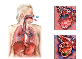
Pneumonia is an infection of the lungs. The lungs are made up of small sacs called alveoli, which fill with air when a healthy person breathes. When an individual has pneumonia, the alveoli are filled with pus and fluid, which makes breathing painful and limits oxygen intake. Many different germs can cause pneumonia, including bacteria, viruses, and fungi.

Causes of Pneumonia

The most common are caused by viruses, including adenoviruses, rhinovirus, influenza virus (flu), respiratory syncytial virus (RSV), and parainfluenza virus (which causes croup).

Symptoms of Pneumonia

The symptoms of pneumonia include:
  • rapid or difficult breathing
  • cough
  • fever
  • chills
  • loss of appetite
  • wheezing (more common in viral infections).
  • nasal congestion
  • breathing with grunting or wheezing sounds
  • vomiting
  • chest pain
  • abdominal pain
  • decreased activity
  • nausea
  • diarrhea
Diagnosis of Pneumonia

Some of these tests may include:
  •     sputum tests (lab tests done on the mucus or phlegm that you cough up from your lungs)
  •     blood tests
  •     chest X-rays
Treatment of Pneumonia

Pneumonia can be treated with antibiotics. These are usually prescribed at a health centre or hospital, but the vast majority of cases of childhood pneumonia can be administered managed effectively within the home. Hospitalization is recommended in infants aged two months and younger, and also in very severe cases.
 

Nursing Plan for Pneumonia
3 Nursing Diagnosis and Interventions for Pneumonia

1. Ineffective airway clearance related to inflammation, secret buildup.

Goal: Effective airway, pulmonary ventilation is adequate and there is no secret buildup.

Nursing Interventions:
  1. Monitor respiratory status every 2 hours, assess the increase in respiratory status and abnormal breath sounds.
  2. Perform percussion, vibration and postural drainage every 4-6 hours.
  3. Give oxygen therapy according to the program.
  4. Help patients cough up secretions / suctioning.
  5. Give a comfortable position that allows the patient to breathe.
  6. Create a comfortable environment so that patients can sleep.
  7. Monitor blood gas analysis to assess respiratory status.
  8. Give drink enough.
  9. Provide sputum for culture / sensitivity test.
  10. Collaboration of antibiotics and other drugs according to the program.

2. Impaired gas exchange related to changes in alveolar capillary membrane.

Goal: Patients showed improved ventilation, optimal gas exchange and tissue oxygenation adequately.

Nursing Interventions:
  1. Observation of level of consciousness, respiratory status, cyanosis signs every 2 hours.
  2. Give Fowler position / semi-Fowler.
  3. Give oxygen according to the program.
  4. Monitor blood gas analysis.
  5. Create an environment that is quiet and patient comfort.
  6. Prevent the occurrence of fatigue in patients.

3. Fluid Volume Deficit related to inadequate oral intake, fever, tachypnoea.

Goal: Patient will maintain normal body fluids.

Nursing Interventions:
  1. Record intake and output of fluids. Encourage the mother to continue giving fluids orally and avoid the condensed milk / drink cold or cough inducing.
  2. Monitor fluid balance in the mucous membranes, skin turgor, rapid pulse, decreased consciousness, vital signs.
  3. Keep drip infusion accuracy according to the program.
  4. Perform oral hygiene.

Milieu Therapy for Low Self-Esteem, Depression and Suicide

Milieu Therapy for Low Self-Esteem, Depression and Suicide
Milieu Therapy has been around since the late 1800's, when the moral care and therapeutic environment to psychiatric nursing major problem. Milieu Therapy can be a powerful therapeutic tool when dynamic individual and social systems can be combined in a planning and meaningful ways to organize and change the behavior and social relations.

Milieu Therapy is a treatment in which the planning of events and interactions every day therapeutic designed with the goal of improving social skills and build self-esteem of patients. Meanwhile, according to Wilson (1992) Mileu Therapy is the use of the environment for therapeutic purposes. Every interaction with a patient is considered to provide beneficial results in improving function optimally.

The Purpose of Milieu Therapy,  include:
  • Improving patient experience positive mental / psychological.
  • Assist individuals in improving self-esteem.
  • Improving the ability to interact with others.
  • Cultivating an attitude trust others.
  • Prepare yourself back into society and achieve positive health changes / optimal.

Characteristics of Milieu Therapy


Characteristics of Milieu Therapy include:
  • familiar
  • cozy
  • physically and psychologically safe
  • ease of access to basic needs
  • staff appreciate the client
  • accept the client's behavior in response to stress
  • respect the rights and opinions of clients
  • right to informed choice
  • easy supervision 24 hours
  • there is a process of information exchange
  • there are socialization, group interaction, and therapeutic communication
  • share responsibility and client engagement

Milieu Therapy for Low Self-Esteem, Depression and Suicide

The physical environment:
  • The room is comfortable and safe.
  • Protected from tools that could be used to injure themselves or others.
  • Medical devices, pharmaceuticals and medical fluid type, locked closet.
  • The room should be placed on the floor of the entire room easily monitored and health workers.
  • The room interesting by placing a bright posters and increase the excitement of life of patients, bright wall colors.
  • The existence of easy reading, humorous and motivating life.
  • Presents cheerful music, TV and movie comedy.
  • Cabinets to store personal belongings of patients.
Social environment:
  • Therapeutic communication, by all officers greet patients as often as possible.
  • Provide an explanation of each will conduct nursing or other medical activities.
  • Receiving the patient's presence.
  • Do not ridicule, belittle.
  • Improving self-esteem of patients
  • Help assess and improve social relationships gradually.
  • Helping patients to interact with his family.
  • Include the family in the plan of nursing
  • Do not leave the patient alone for too long.

Home Care - Prevent and Treat Vaginal Discharge

Prevent and Treat Vaginal Discharge
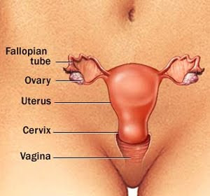
There are some women whose vaginal discharge is so light that they don’t realize they even have a discharge. ust like any other physical characteristic, the amount of normal discharge varies a lot from woman to woman.

Vaginal discharge is a term given to biological fluids contained within or expelled from the vagina.

Normal vaginal discharge has several purposes: cleaning and moistening the vagina, and helping to prevent and fight infections. Although it's normal for the color, texture, and amount of vaginal fluids to vary throughout a girl's menstrual cycle, some changes in discharge may indicate a problem.

Vaginal discharge is made up of cervical mucous, discharge from vaginal walls, and sweat from the vulva. Some women have large numbers of mucous glands on the cervix, which increases the amount of vaginal discharge. Overweight women, especially if they wear stretch pants, will have a heavier vaginal discharge because of the increase in sweat.

The following situations can increase the amount of normal vaginal discharge:
  • Emotional stress
  • Ovulation (the production and release of an egg from your ovary in the middle of your menstrual cycle)
  • Pregnancy
  • Sexual excitement

To help prevent and treat vaginal discharge:

  • Keep your genital area clean and dry.
  • Do not douche. While many women feel cleaner if they douche after menstruation or intercourse, it may actually worsen vaginal discharge because it removes healthy bacteria lining the vagina that are there to protect you from infection. It can also lead to infection in the uterus and fallopian tubes, and is never recommended.
  • Use an over-the-counter yeast infection treatment cream or vaginal suppository, if you know that you have a yeast infection.
  • Eat yogurt with live cultures or take Lactobacillus acidophilus tablets when you are on antibiotics to avoid a yeast infection.
  • Use condoms to avoid catching or spreading STIs.
  • Avoid using feminine hygiene sprays, fragrances, or powders in the genital area.
  • Avoid wearing extremely tight-fitting pants or shorts, which may cause irritation.
  • Wear cotton underwear or cotton-crotch pantyhose. Avoid underwear made of silk or nylon, because these materials are not very absorbent and restrict air flow. This can increase sweating in the genital area, which can cause irritation.
  • Use pads and not tampons.
  • Keep your blood sugar levels under good control if you have diabetes.
If the discharge is caused by a sexually transmitted disease, your sexual partner (or partners) must be treated as well, even if they have no symptoms. Failure of partners to accept treatment can cause the infection to keep coming back and may lead to pelvic inflammatory disease or infertility. Source : www.nlm.nih.gov

Nursing Care Plan for Prostate Cancer - 3 Diagnosis and Interventions

Nursing Care Plan for Prostate Cancer - 3 Diagnosis and Interventions
The word "prostate" comes from Medieval Latin prostate and Medieval French prostate. The ancient Greek word prostates means "one standing in front", from proistanai meaning "set before".

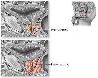
The prostate is an exocrine gland of the male reproductive system, and exists directly under the bladder, in front of the rectum.

Prostate cancer generally affects men over 50 and is rare in younger men. It’s the most common type of cancer in men. Around 37,000 men in the UK are diagnosed with prostate cancer each year.

Prostate cancer is a disease which only affects men. Cancer begins to grow in the prostate - a gland in the male reproductive system. In the vast majority of cases, the prostate cancer starts in the gland cells - this is called adenocarcinoma. In this article, prostate cancer refers just to adenocarcinoma. Prostate cancer is mostly a very slow progressing disease. In fact, many men die of old age, without ever knowing they had prostate cancer - it is only when an autopsy is done that doctors know it was there.

The symptoms of both benign enlargement of the prostate gland and malignant tumours (cancer) are similar and can include any of the following:
  •     difficulty passing urine
  •     passing urine more frequently than usual, especially at night
  •     pain when passing urine
  •     blood in the urine (this is not common).
Nobody is really sure of what the specific causes are. There are so many possible factors, including :
  • Age
  • Genetics
  • Diet
  • Medication
  • Obesity
  • Sexually transmitted diseases (STDs)
  • Agent Orange

3 Nursing Diagnosis and Nursing Interventions for Prostate Cancer


1. Impaired Urinary Elimination related to an enlarged prostate, and bladder distension.

Intervention:
  •     Encourage the patient to urinate every 2-4 hours and when it suddenly felt.
  •     Observation of the flow of urine, note the size and strength.
  •     Percussion / palpation of the suprapubic area.
  •     Encourage fluid intake to 3000 ml per day.
  •     Monitor vital signs closely
  •     Collaboration in the provision of drugs.

2. Resti for Infection related to invasive procedures (tools during surgery)

Intervention:
  • Maintain a sterile catheter system, provide catheter care and give regular antibiotic ointment around the catheter.
  • Perform ambulation with dependent drainage bag.
  • Observation of wound drainage around suprapubic catheter.
  • Replace dressings with frequent (supra incision / retropubic and perineal), cleaning and drying of the skin over time.
  • Collaboration in the provision of antibiotics.

3. Imbalanced Nutrition, Less Than Body Requirements related to the nausea and weight loss

Intervention:
  •      Assess the patient's nutritional status.
  •      Encourage the patient to eat small amounts frequently.
  •      Collaborate with a nutritionist.
  •      Collaborate with the physician in the delivery of antiemetic drugs.

Diabetes Mellitus - 6 Nanda Nursing Diagnosis

Diabetes mellitus is a condition in which the pancreas no longer produces enough insulin or cells stop responding to the insulin that is produced, so that glucose in the blood cannot be absorbed into the cells of the body.

Diabetes mellitus is a chronic disease that causes serious health complications including renal (kidney) failure, heart disease, stroke, and blindness.

Risk factors for type 2 diabetes mellitus are greater for some ethnicities, as mentioned before. Furthermore, those people who have a family history of type 2 diabetes, who are overweight or inactive also face a greater risk of type 2 diabetes mellitus.

Diabetes mellitus affects a variety of people of all races, ages and nations. It is unkown why some people develop type 1 diabetes.

It may be linked to environmental factors or a virus however it has been estabilished if there is a family history of type 1 diabetes then there is a higher risk of developing type 1 diabetes.

Symptoms include frequent urination, lethargy, excessive thirst, and hunger. The treatment includes changes in diet, oral medications, and in some cases, daily injections of insulin.

Symptoms of diabetes can develop suddenly (over days or weeks) in previously healthy children or adolescents, or can develop gradually (over several years) in overweight adults over the age of 40. The classic symptoms include feeling tired and sick, frequent urination, excessive thirst, excessive hunger, and weight loss.

Diabetes is suspected based on symptoms. Urine tests and blood tests can be used to confirm a diagnose of diabetes based on the amount of glucose found. Urine can also detect ketones and protein in the urine that may help diagnose diabetes and assess how well the kidneys are functioning. These tests also can be used to monitor the disease once the patient is on a standardized diet, oral medications, or insulin.

Research continues on diabetes prevention and improved detection of those at risk for developing diabetes. While the onset of Type I diabetes is unpredictable, the risk of developing Type II diabetes can be reduced by maintaining ideal weight and exercising regularly. The physical and emotional stress of surgery, illness, pregnancy, and alcoholism can increase the risks of diabetes, so maintaining a healthy lifestyle is critical to preventing the onset of Type II diabetes and preventing further complications of the disease.

There is currently no cure for diabetes. The condition, however, can be managed so that patients can live a relatively normal life. Treatment of diabetes focuses on two goals: keeping blood glucose within normal range and preventing the development of long-term complications. Careful monitoring of diet, exercise, and blood glucose levels are as important as the use of insulin or oral medications in preventing complications of diabetes. In 2003, the American Diabetes Association updated its Standards of Care for the management of diabetes. These standards help manage health care providers in the most recent recommendations for diagnosis and treatment of the disease.

Diabetes Mellitus - 6 Nanda Nursing Diagnosis

1. Fluid Volume Deficit

related to:
osmotic diuresis (hyperglycemia).

2. Imbalanced Nutrition, Less Than Body Requirements

related to
poor nutrition intake.

3. Risk for Infection

related to:
high glucose levels
reduction in leukocyte function.

4. Knowledge Deficit: about the disease process

related tyo: lack of information.

5. Risk for Impaired Skin Integrity

related to:
immobilization
neuropathy.

6. Activity Intolerance

related to:
physical weakness.

9 Sample of Pulmonary Tuberculosis (PTB) Nursing Diagnosis - Nanda

pulmonary tuberculosis nanda nursing diagnosis
Tuberculosis (TB) is an infectious disease that is caused by a bacterium called Mycobacterium tuberculosis.

Symptoms of TB in the lungs may include
  • A bad cough that lasts 3 weeks or longer
  • Weight loss
  • Coughing up blood or mucus
  • Weakness or fatigue
  • Fever and chills
  • Night sweats
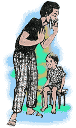
Tuberculosis is ultimately caused by the Mycobacterium tuberculosis that is spread from person to person through airborne particles. It is not guaranteed, though, that you will become infected with TB if you inhale the infected particles. Some people have strong enough immune systems that quickly destroy the bacteria once they enter the body. Others will develop latent TB infection and will carry the bacteria but will not be contagious and will not present symptoms. Still others will become immediately sick and will also be contagious. 

9 Sample of Pulmonary Tuberculosis (PTB) Nursing Diagnosis - Nanda

1. Impaired Gas Exchange

related to:
  • the exudate in alveo
  • surface of the lung function decline.

2. Ineffective Airway Clearance

related to:
  • increased sputum
  • reduction effort to cough.

3. Ineffective Breathing Pattern

related to
  • inflammation
  • fatigue.

4. Hyperthermia

related to:
  • the infection process.

5. Fluid Volume Deficit

related to:
  • fever
  • fatigue due to lack of fluid intake.

6. Imbalanced Nutrition, Less Than Body Requirements

related to:
  • decreased appetite,
  • fatigue
  • dyspnea.

7. Risk for [spread] of Infection

related to:
  • lower resistance of others who are around people.

8. Ineffective management Therapeutic regimen

related to:
  • lack of knowledge about the disease process.

9. Activity Intolerance

related to:
  • fatigue,
  • changes in nutritional status
  • fever.

Airway Management of Tuberculosis

Nursing Care Plan for Tuberculosis
Nursing Diagnosis Interventions for Tuberculosis

Pulmonary tuberculosis (TB) is caused by the bacteria Mycobacterium tuberculosis (M. tuberculosis). M. tuberculosis is an aerobic, nonmotile, non-spore-forming rod that is highly resistant to drying, acid, and alcohol.

The probability of transmission from one person to another depends on the number of infectious droplets expelled by a carrier, the duration of exposure, and the virulence of the M. tuberculosis.

Most people who develop symptoms of a TB infection first became infected in the past. However, in some cases, the disease may become active within weeks after the primary infection.

The following people are at higher risk for active TB:
  •     Elderly
  •     Infants
  •     People with weakened immune systems, for example due to AIDS, chemotherapy, diabetes, or certain medications.
The following factors may increase the rate of TB infection in a population:
  •     Increase in HIV infections
  •     Increase in number of homeless people (poor environment and nutrition)
  •     The appearance of drug-resistant strains of TB

A definitive diagnosis of TB can only be made by culturing M. tuberculosis organisms from a specimen taken from the patient. However, TB can be a difficult disease to diagnose, mainly because of the difficulty in culturing this slow-growing organism in the laboratory. A complete evaluation for TB must include a medical history, a chest radiograph, a physical examination, and microbiologic smears and cultures. It may also include a tuberculin skin test and a serologic test.

Airway Management of Tuberculosis


The most common cause of inflammatory stricture of the bronchus is TB. Tracheobronchial TB has been reported in 10–20% of all patients with pulmonary TB. The principal CT findings of airway TB are circumferential wall thickening and luminal narrowing, with involvement of a long segment of the bronchi. In active disease, the airways are irregularly narrowed in their lumina and have thick walls, whereas in fibrotic disease, the airways are smoothly narrowed and have thin wall. The left main bronchus is involved more frequently in fibrotic disease, whereas both main bronchi are equally involved in active disease.


Nursing Care Plan for Tuberculosis

Nursing Diagnosis : Ineffective Airway Clearance related to increased efforts to decrease sputum and cough.

Goal :: Improve cleanliness is to decrease airway secretions and repair ien to cough.

Intervention:

1. Encourage clients to drink 8 glasses of water / 2 liters of water a day (other than milk) for dilution secretion, while milk may increase the secretion.
R / Reassure clients that water moisturizes breathing.

2. Reassure clients that water moisturizes breathing.
R / Humidity helps airway secretions and allowing greater.

3. Encourage clients to cough effectively and breathe deeply.
R / proper coughing technique, a way to remove the sputum.

4. Encourage clients to rest between intervals cough and to change positions every 12 hours when possible.
R / Rest and changing position helps to reduce fatigue and overall spending sputum, insert oxygen to regenerate the cells.

5. Explain to the client's intended use of expectorant if found.
R / Expectorants help to loosen airway secretions and expenses.

6. Observations sputum characteristics coming out, discoloration, odor consistency / amount. Report immediately if there is a change.
R / normal sputum is thin and translucent white when mixed with blood, may indicate purulent complications.

Risk for Injury - Alzheimer's Disease Nursing Care Plan

Alzheimer's disease is a brain disease that slowly destroys memory and thinking skills and, eventually, the ability to carry out the simplest tasks. People may have trouble remembering things that happened recently or names of people they know. Memory problems are one of the first signs of Alzheimer's. Over time, symptoms will most often get worse, and problems can include getting lost, repeating questions, and taking longer than normal to finish daily tasks. As the disease progresses, people may have trouble learning new things, recognizing family and friends, and communicating. Eventually, they need total care.

Alzheimer's disease is named after Dr. Alois Alzheimer, a German doctor. In 1906, Dr. Alzheimer noticed changes in the brain tissue of a woman who had died of an unusual mental illness. He found many abnormal clumps (now called amyloid plaques) and tangled bundles of fibers (now called neurofibrillary tangles). Today, these plaques and tangles in the brain are considered the main signs of Alzheimer's disease.

People with Alzheimer's disease are at a serious disadvantage. Their impairments in memory and reasoning severely limit their ability to act appropriately in crises.
Specific home safety precautions may apply and environmental changes may be needed.

Prevention begins with a safety check of every room in your home. Use the following room-by-room checklist to alert you to potential hazards and to record any changes you need to make. You can buy products or gadgets necessary for home safety at stores carrying hardware, electronics, medical supplies, and children's items.

Risk for Injury - Alzheimer's Disease Nursing Care Plan

Nursing Care Plan for Alzheimer's Disease - Risk for Injury

Nursing Diagnosis : Risk for Injury
related to:
  •     Unable to recognize / identify hazards in the environment.
  •     Disorientation, confusion, impaired decision making.
  •     Weakness, the muscles are not coordinated, the presence of seizure activity.

Nursing Interventions and Rational :


Nursing Intervention

  1. Assess the degree of impaired ability of competence emergence of impulsive behavior and a decrease in visual perception.
  2. Help the people closest to identify the risk of hazards that may arise.
  3. Eliminate / minimize sources of hazards in the environment
  4. Divert attention to a client when agitated or dangerous behaviors like getting out of bed by climbing the fence bed.

Rational:
  1. Impairment of visual perception increase the risk of falling. Identify potential risks in the environment and heighten awareness so that caregivers more aware of the danger.
  2. An impaired cognitive and perceptual disorders are beginning to experience the trauma as a result of the inability to take responsibility for basic security capabilities, or evaluating a particular situation.
  3. Maintain security by avoiding a confrontation that could improve the behavior / increase the risk for injury.

10 NANDA COPD Chronic Obstructive Pulmonary Disease

Chronic obstructive pulmonary disease (COPD) is a general term which includes the conditions chronic bronchitis and emphysema. COPD is the preferred term, but you may still hear it called chronic obstructive airways disease (COAD).
  •     Chronic means persistent.
  •     Bronchitis is inflammation of the bronchi (the airways of the lungs).
  •     Emphysema is damage to the smaller airways and air sacs (alveoli) of the lungs.
  •     Pulmonary means 'affecting the lungs'.
The term COPD is used to describe airflow obstruction due to chronic bronchitis, emphysema, or both.

The sudden risk caused by COPD is due to the increase in people who smoke and the demographic changes in many countries. In the US, COPD is considered as the fourth leading cause of death. In economic terms the cost of the disease to the US economy in 2007 is pegged at $42.6 billion in terms of health care costs and loss in productivity.

The symptoms of COPD include: constant cough; excess sputum (mucus) production; shortness of breath while doing activities you used to be able to do; wheezing, or whistling sound when you breathe; and tightness in the chest.

The most common symptoms of COPD are breathlessness, or a 'need for air', excessive sputum production, and a chronic cough. However, COPD is not just simply a "smoker's cough", but a under-diagnosed, life threatening lung disease that may progressively lead to death.

The loss of lung function in COPD patients is so gradual that many patients do not realize that they have the condition until it is severe. By the time most patients seek medical attention, they may have lost 50% of their pulmonary function.

There is a need for greater awareness of COPD and early diagnosis and treatment can retard progression of disease and improve quality of life. A person who has COPD should adopt a number of strategies in order to manage and to combat this lung disease. Some of these important strategies include saying no to smoking, vaccinations, rehabilitation and drug therapy. Drug therapies can be done thru the use of inhalers.

The inhalers that are suggested help dilate the airways and the theophylline. Most of the time, the inhaled steroids can be used to contain lung inflammation and can suppress flare-ups. Usually antibiotics are also used during the flare-ups of the symptoms of COPD.

Nursing Diagnosis COPD Care Plan


10 NANDA - Nursing Diagnosis for COPD Chronic Obstructive Pulmonary Disease 
  1. Ineffective airway clearance 
  2. Ineffective breathing pattern 
  3. Impaired gas exchange 
  4. Activity intolerance
  5. Imbalanced Nutrition: less than body requirements
  6. Disturbed sleep pattern
  7. Bathing / Hygiene Self-care deficit 
  8. Anxiety 
  9. Ineffective individual coping 
  10. Deficient Knowledge

Nursing Care Plan for Goiter - Assessment and Diagnosis

Nursing Assessment and Nursing Diagnosis for Goiter

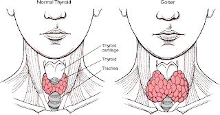
Goiter (struma), is a swelling of the thyroid gland, which can lead to a swelling of the neck or larynx (voice box). Goitre is a term that refers to an enlargement of the thyroid (thyromegaly) and can be associated with a thyroid gland that is functioning properly or not.

A person with goiter can have normal levels of thyroid hormone (euthyroidism), excessive levels (hyperthyroidism) or levels that are too low (hypothyroidism).

A symptom is something the patient feels or reports, while a sign is something other people, including the doctor detects. For example, a headache may be a symptom while a rash may be a sign.

Some patients may have goiter and not know it because they have no symptoms.

The main symptom for a person with goiter is swelling of the thyroid gland. This may eventually become a noticeable lump in the throat. The patient may be more aware of it - a visible swelling at the base of the neck - when looking in the mirror and shaving or putting on makeup.

The following symptoms may also exist when a person has goiter:
  • Hoarseness (voice)
  • Coughing more frequently than usual
  • A feeling of tightness in the throat
  • Swallowing difficulties (less common)
  • Breathing difficulties (less common)
Nursing Care Plan for Goiter

Nursing Care Plan for Goiter - Assessment and Diagnosis

In implementing the nursing care, the authors use the guidelines as a basis for solving the nursing care of patient problems scientifically and systematically, which includes the step of assessment, nursing diagnosis, nursing Interventions and evaluation.


Assessment

Assessment is the first step in the nursing process as a whole in order to get the data or information needed to determine the health problems faced by patients through interviews, observation, and physical examination include:

a. Activity / rest
Subjective data: insomnia, muscle weakness, impaired coordination, severe fatigue.
Objective data: muscle atrophy.

b. Elimination
Subjective data: urine in large amounts, changes in the faeces, diarrhea.

c. Ego integrity
Subjective data: experiencing severe stress both emotionally and physically.
Objective data: emotional instability, depression.

d. Food / fluid
Subjective data: sudden weight loss, increased appetite, eat a lot, eat often, thirst, nausea and vomiting.
Objective data: thyroid enlargement, goiter.

e. The pain / comfort
Subjective data: orbital pain, photophobia.

f. Breathing
Subjective data: increased respiratory frequency, tachypnea, dyspnea, pulmonary edema (the crisis thyrotoxicosis).

g. Security
Subjective data: intolerance to heat, excessive sweating, allergic to iodine (may be used in the examination).
Objective data: the temperature rises above 37.40 C, diaphoresis, smooth skin, warm and reddish, thin hair, shiny and straight, eksoptamus: retraction, conjunctival irritation and watery, pruritus, erythema lesion (common in pretibial) is a very severe.

h. Sexuality
Data subyktif: decreased libido, bleeding slightly or not at all, impotence.


After all the data collected, further divided into two groups:

a. subjective data
Subjective data include: coordination disorder insomnia, changes in the pattern of elimination, the ability to handle the pressure (stress), weight loss, increased appetite, orbital pain, respiratory frequency increases, the power adjustment to heat and cold, decreased libido.

b. objective data
It is characterized by muscle atrophy, emotional instability, depression, thyroid enlargement, goiter, increased temperatures above 37.40 C, diaphoresis, nature and characteristics of the body, including hair quality situation and the state of the eye.


The next step is determining the nursing diagnosis is a statement and a real or potential problem, based on the data collected.


Nursing Diagnosis for Goiter

Nursing diagnosis in patients with goitre especially post surgery can be formulated as follows:

1. Risk for Ineffective Airway Clearance related to obstruction of the trachea, swelling, bleeding and laryngeal spasm,
characterized by:
Subjective data: pain swallowing, painful wound.
Objective data: breathing fast and deep, there are secretions / mucus.

2. Impaired Verbal Communication related to vocal cord injury / damage to the larynx, tissue edema, pain, discomfort,
characterized by:
Subjective data: swelling of the throat tissues, pain in the wound, the patient does not feel comfortable, pain swallowing.

3. Risk for Injury / tetany related to the surgery, stimulation of the central nervous system,
characterized by:
Subjective data: rapid breathing (tachypnea), wound pain.
Objective data: increased body temperature, tachycardia, cyanosis, convulsions, numbness, and infection of the surgical wound.

4. Acute Pain related to the surgery of the tissue / muscle and postoperative edema,
characterized by:
Subjective data: ask, ask for information, statements misconceptions.
Objective data: do not follow the instructions / complications that can be prevented.

CVA - Stroke Definition and Nursing Diagnosis

CVA - Stroke
A cerebral vascular accident is another name for a stroke. It is damage to the brain caused by a disruption of the blood supply to a part of the brain. This disruption of blood supply can be caused by a blood clot, or by a ruptured artery.

The symptoms of a cerebral vascular accident depend on which part of the brain is affected. Common symptoms may include paralysis of a part of the body, loss of all or part of the vision, or loss of the ability to speak or to understand speech.

A stroke is a medical emergency and can cause permanent neurological damage, complications, and death. Risk factors for stroke include old age, high blood pressure, previous stroke or transient ischemic attack (TIA), diabetes, high cholesterol, tobacco smoking and atrial fibrillation. High blood pressure is the most important modifiable risk factor of stroke. It is the second leading cause of death worldwide.

Stroke symptoms typically start suddenly, over seconds to minutes, and in most cases do not progress further. The symptoms depend on the area of the brain affected. The more extensive the area of brain affected, the more functions that are likely to be lost. Some forms of stroke can cause additional symptoms. For example, in intracranial hemorrhage, the affected area may compress other structures. Most forms of stroke are not associated with headache, apart from subarachnoid hemorrhage and cerebral venous thrombosis and occasionally intracerebral hemorrhage.

Stroke is diagnosed through several techniques: a neurological examination (such as the Nihss), CT scans (most often without contrast enhancements) or MRI scans, Doppler ultrasound, and arteriography. The diagnosis of stroke itself is clinical, with assistance from the imaging techniques. Imaging techniques also assist in determining the subtypes and cause of stroke. There is yet no commonly used blood test for the stroke diagnosis itself, though blood tests may be of help in finding out the likely cause of stroke.

Nursing Diagnosis for CVA - Stroke:

1. Ineffective Cerebral Tissue Perfusion

2. Impaired Physical Mobility.

3. Imbalanced Nutrition, Less Than Body Requirements.

4. Impaired Skin Integrity.

5. Impaired Verbal Communication

6. Disturbed Sensory Perception

7. Self-Care Deficit

8. Knowledge Deficit

NANDA Nursing

Nursing Care Plan